
Labs and Activities
Week 5 – Sight, Hearing, and Equilibrium
Learning Objectives
Upon completion of this learning package, the student will be expected to recall the following.
- Describe the pathway of taste and smell sensations into its area of interpretation in the cerebral cortex considering for each:
- stimuli activating receptors
- location, structure and response of receptors
- specific sensory nerve pathway(s) to the area of interpretation
- specific area of interpretation in cerebral cortex
Click the triangle drop-down to see specific objectives:
Photoreceptors: Sight
- Recognize that the eyeball is divided into three layers (coats):
- the outer fibrous tunic
- the middle vascular tunic
- the inner retina tunic
- State the location (using provided diagrams), the structural characteristics, and the function(s) of the following structures which describe the outer layer of the eye:
- sclera
- cornea
- canal of Schlemm
- State the location (using provided diagrams), the structural characteristics, and the function(s) of the following structures which form the middle layer of the eye:
- choroid
- ciliary body
- iris (pupil)
- State the location, structural characteristics and the function in the physiology of vision for each of the following structures that form the inner layer of the eye:
- retina
- rods
- cones
- fovea centralis (central fovea)
- optic disc
- State, considering the lens of the eye:
- the function of the zonular fibres (suspensory ligaments)
- the function and type of innervation of the ciliary muscles
- the relevance of the lens shape to vision
- the function of the lens in terms of refraction and image formation
- State, considering the two cavities of the interior of the eyeball (anterior cavity and posterior cavity:
- the two divisions of the anterior cavity and their location
- the name and consistency of fluid in each cavity
- the origin and cycling of fluid in the anterior cavity
- the function of each fluid
- Identify for both extrinsic and intrinsic eye muscles:
- kind of muscle tissue
- type of movement
- specific functions
- Identify the function of the following external structures associated with the eye:
- eyebrow and eyelashes
- eyelids
- orbit wall
- State, considering the lacrimal apparatus:
- its composition -5 structures
- its function in protection of the eye
- Explain how the process occurs and why it contributes to visual acuity of near objects, for each of the following processes necessary for visual acuity of near objects:
- accommodation of lens
- pupil constriction
- convergence of the eyeballs
Mechanoreceptors: Hearing
- Identify and locate each of the following components of the ear involved in responding to the presence of sound: (Use provided diagrams)
- External ear
- auricle (pinna)
- external auditory canal (acoustic meatus)
- tympanic membrane
- Middle ear
- malleus
- incus
- stapes
- tympanic cavity
- Inner ear
- cochlea (part of the bony labyrinth)
- oval window
- cochlear duct
- perilymph
- endolymph
- spiral organ (of Corti)
- round window
- External ear
- State the functions of the following components of the ear in hearing:
- auricle (pinna)
- external auditory canal (acoustic meatus)
- tympanic membrane
- ossicles (malleus, incus, stapes)
- oval window
- perilymph (inside cochlea)
- endolymph (inside cochlear duct)
- tectorial membrane
- basilar membrane
- round window
- State the function of the following receptor structure of the inner ear in the sensation of hearing:
- spiral organ (organ of Corti)
- State the location and function in relation to hearing for each of the following components:
- temporal bone
- auditory (Eustachian) tube
Mechanoreceptors: Equilibrium
- Identify and locate each of the following components of the ear involved in responding to changes in body/head positions and/or velocity: (Use provided diagrams)
- From the inner ear –bony labyrinth
- a) vestibule
- b) semicircular canals
- From the inner ear –membranous labyrinth
- c) utricle
- d) saccule
- e) semicircular ducts
- From the inner ear –bony labyrinth
- Define the term “vestibular apparatus”
Lab Activity 1: Dissection of an Eye
Materials Required
- Dissecting tray and instruments
- Preserved cow eye
- Eye model
- Functioning eye model
- Gloves
Part 1A: Dissection
- Obtain a pair of gloves, a dissecting tray and a set of dissecting instruments.
- Obtain a preserved eye from the lab instructor.
- Trim away fat and other connective tissue (if present). Be careful not to cut the eyeball.
- Examine the surface of the eyeball carefully. Locate the optic nerve (if present), and the extrinsic muscles.
Question: What kind of muscles are the extrinsic eye muscles?
Question: What functions do the extrinsic eye muscles serve?
Question: What is the branch of the efferent nervous system that goes to the extrinsic eye muscles?
- Obtain a model of the eye and locate the six extrinsic eye muscles.
Question: What are the names of the extrinsic eye muscles? As you name each muscle move the eye model as it would if that muscle were contracting. At the same time move your own eye the same way.
- Now look at the front of preserved eye and note the shape of the pupil if it is visible. Is it round or elliptical?
- Note the conjunctiva which lines the eyelid and is reflected over the anterior surface of the eye. Lift some of this thin membrane away from the eye with forceps and examine it.
Question: What is the name of this tunic?
Question: How is the anterior part of this coat modified when it overlies the coloured part of the eye?
Question: What is the anterior portion of the outer coat of the eyeball called
- If you have removed all of the fat you will have exposed the outermost tunic of the eyeball.
Question: What part of the eye is covered with the conjunctiva?
- Holding the eyeball firmly, but gently, with a sharp scalpel make an incision in the sclera about 1/4” posterior to the edge of the cornea. (NOTE how difficult it is to penetrate this tissue).
- Insert scissors into this opening. Holding the scissors parallel to the eyeball wall, cut completely around the cornea.
Be careful not to squeeze the eyeball during this procedure to avoid loss of fluid from the eye.
- Gently separate the anterior portion of the eyeball from the posterior portion. Place the anterior portion on your tray with the inner surface upward.
Note: the lens usually remains attached to the vitreous body, however, it may be attached to the anterior portion of the eye.
- Looking at the inner surface of the anterior portion of the eyeball and identify:
- the thick, black, circular ciliary body.
- the iris
- Now examine the iris.
Question: What is the function of the iris?
Question: What type of tissue is the iris and how is it arranged?
Question: What will happen to your pupil when:
- the circular fibres contract?
- radial fibres contract?
Question: Which branch of the efferent nervous system goes to the iris?
- Now examine the ciliary body. Using the following instructions on the following pages, use the functioning eye model to see how the lens affects focusing of the image. You may do this now or you may complete the dissection first before proceeding to the eye model.
Instructions for the Functioning Eye Model
- Turn the light on and place the letter stand with the red letter Y at the 10cm mark of the meter stick.
- Make sure that the adjustment nut on top of the eye model is lined up beside the center of the 3 circles.
- Use the syringe at the back of the eye model to get the image in focus on the retina. (Note: the retina can be moved up and down to center the image.)
Question: How does the image on the retina appear relative to the letter on the stand?
- Move the letter stand to the 25cm mark.
Question: What happens to the image on the retina?
- Use the syringe to increase the amount of water in the lens.
Question: What happens to the shape of the lens?
Question: What does changing the shape of the lens do to the image on the retina?
- Return the letter stand to the 10 cm mark and use the syringe to readjust the focus.
- Adjust the eye model so that the adjustment nut on top of the model is lined up beside the posterior circle.
Question: What does this do to the image on the retina?
- Place the +2.00 lens in the lens holder in front of the eye model.
Question: What happens to the image on the retina?
- Remove the lens and place it back in the envelope.
- Now adjust the eye model so that the adjustment nut is beside the anterior circle.
Question: What happens to the image on the retina?
- Put the -2.00 lens in the lens holder.
Question: What happens to the image on the retina?
- Replace the -2.00 lens with the + 2.00 lens .
Question: What happens to the image on the retina?
- When you have finished turn off the light and make sure that you return the +2.00 and -2.00 lenses to the correct envelopes.
Dissection Continued
- Look to see if there is any aqueous humor left between the cornea and iris.
Question: How does the consistency of aqueous humor compare to the consistency of the vitreous body?
- Now examine the posterior portion of the eye. Note the vitreous humor (body). This jelly- like mass helps to hold the lens in place anteriorly, and holds the retina against the choroid coat.
- Carefully invert the eyeball and remove the vitreous body.
- After you have removed the vitreous body, examine the inner surface of the eye and locate the layer of yellow tissue. This layer is very thin and some of it may have torn off when you removed the vitreous body.
- Using a blunt probe spread this layer out flat on the inner surface of the eye. (It may help to place the eye in a beaker of water).
Question: What is the name of this layer?
Question: What type of tissue is this layer?
Question: What is the function of this layer?
Question: Why is there no macula lutea in a cow eye?
- Using a blunt probe gently lift the yellow layer to expose the iridescent layer underneath.
- Using a probe lift this layer off of the underlying layer.
Question: What type of tissue is this layer?
Question: How is this layer different from the yellow layer?
Question: What are the functions of this layer?
Question: Where is the yellow layer attached to this underlying layer?
- Using your finger touch the blue layer and note the brown pigment that comes off on your finger.
Question: What is the function of this pigment?
- Using a probe lift the blue layer up to expose the next layer.
Question: What is the name of the layer exposed by this procedure?
- Note the thickness of this layer as compared to the first two.
Question: What type of tissue is this layer?
Question: What are the functions of this layer besides attachment of extrinsic muscles?
- Examine the optic nerve.
Question: What is the number of this cranial nerve?
Question: What colour is the optic nerve? Explain.
Question: Is this nerve afferent or efferent in nature? Explain.
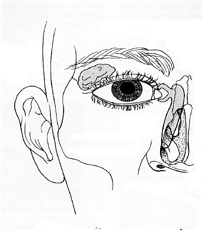
- On the diagram below trace the path of lacrimal fluid naming the parts of the lacrimal apparatus. (Hint: use the diagram available in the lab)
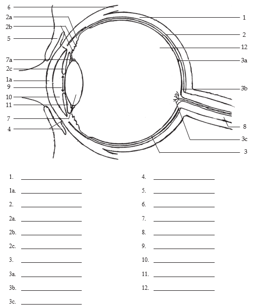
- Download and label the following figure (5.2) by right-clicking on the image and choosing “Save Image As”.
After you have completed your lab activities dispose of the dissected eye in the Biohazard Container and rinse off your dissecting instruments.
Review – 1A
Download Structural Composition and Functional Components of the Eye [DOCX] to print and fill out.
For each of the following components of the eye (Table 5.1 Structural Composition and Functional Components of the Eye 1-10) involved in the transmission of light and/or protection of the eye:
- select the most appropriate structural composition
- Mucous membrane covering anterior surface of eyeball
- Receptors for dim light
- Smooth, sphincter muscle
- Transparent anterior part of sclera
- Clear fluid filling anterior cavity
- Composed of white, fibrous tissue
- Voluntary muscle attached to bony orbit and superior part of eyeball
- Area of retina containing many cones
- Smooth muscle at anterior aspect of choroid
- Vascular, pigmented layer
- select the most appropriate function
- Regulates amount of light entering eyeball
- Maintains intraocular pressure
- Rolls eyeball upward
- Changes the shape of the lens
- Provides nutrients to retina and sclera
- Keeps cornea moist
- Provides shape for the eyeball
- Discriminate between different shades of dark and light
- Lets light into eye
- Sharpest point of vision on retina
| Component | Structural Composition | Function |
|---|---|---|
| 1. Conjunctiva | ||
| 2. Cornea | ||
| 3. Iris | ||
| 4. Sclera | ||
| 5. Choroid | ||
| 6. Rods | ||
| 7. Fovea centralis | ||
| 8. Aqueous humour | ||
| 9. Ciliary muscle | ||
| 10. Superior rectus muscle |
Lab Activity 2: the Ear
Materials Required:
- Diagrams for labelling
Part 2A – The Ear: Structures of the Ear
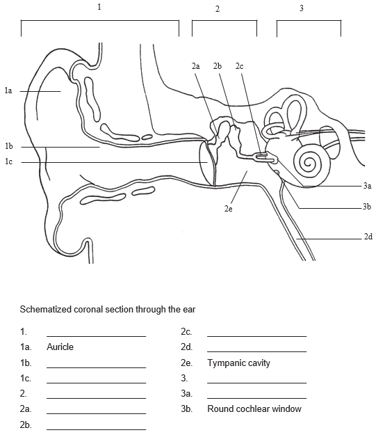
- Download and label the following figure (5.3) by right-clicking on the image and choosing “Save Image As”.
Review – 2A
Download Structural Composition and Functional Components of the Ear [DOCX] to print and fill out.
For each of the following components of the ear (Table 5.2 Structural Composition and Functional Components of the Ear (1-6) involved in the transmission of sound:
- select the most appropriate structural composition
- Wall consists of bone lined with cartilage
- Small opening between middle and inner ear
- Small bones extending across middle ear
- Contains hair cells projecting into fluid
- Semitransparent partition of fibrous connective tissue
- Fluid surrounding the membranous cochlear duct
- select the most appropriate function
- Conducts sound waves from external acoustic meatus to ossicles
- Causes the cochlear duct to vibrate
- Relays sound waves from ossicles to perilymph
- Conducts sound waves to tympanic membrane
- Receptors for auditory sensations
- Relay sound waves to oval window
| Component | Structural Composition | Function |
|---|---|---|
| 1. External acoustic means | ||
| 2. Tympanic membrane | ||
| 3. Ossicles | ||
| 4. Oval window | ||
| 5. Perilymph | ||
| 6. Organ of Corti |
Part 2B – The Ear: Structural Components of the Ear Involved in Equilibrium
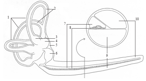
- Download and label the following figure (5.4) by right-clicking on the image and choosing “Save Image As”.
- semicircular canals
- semicircular ducts
- ampulla of semicircular duct
- utricle
- vestibule
- saccule
- cochlear duct
- spiral organ of Corti
- scala tympani
- scala vestibuli
Review – 2B
Download Structural Composition and Functional Components of the Ear Involved in Equilibrium [DOCX] to print and fill out.
For each of the following components of the ear (Table 5.3 Structural Composition and Functional Components of the Ear Involved in Equilibrium (1-5) involved in the transmission of sound:
- select the most appropriate structural composition
- Bony structure containing two sac-like structures
- Three membranous ducts arranged at right angles to each other
- Contain sensory hair cells coated with a gelatinous layer which contains otoliths
- Three bony structures arranged at right angles to each other
- Hair cells covered with gelatinous material located within semi-circular ducts
- select the most appropriate function
- Sense organs of static equilibrium
- Sense organs of dynamic equilibrium
- Contains endolymph which stimulates hair cell
- Contains perilymph surrounding utricle and saccule
- Contains membranous ducts
| Component | Structural Composition | Function |
|---|---|---|
| 1. Vestibule | ||
| 2. Utricle and saccule | ||
| 3. Semicircular canals | ||
| 4. Semicircular ducts | ||
| 5. Hair cells on cristae |
