
Labs and Activities
Week 4 – The Cerebrum
Learning Objectives
Upon completion of this learning package, the student will be expected to recall the following.
Click the triangle drop-down to see specific objectives:
The Cerebrum
- Describe the gross anatomy of the cerebrum as follows:
- location
- number of hemispheres
- name of the fissure and associated tissues dividing the hemispheres
- name and number of lobes per hemisphere
- general distribution of grey and white matter
- descriptive terms related to the surface: gyri; fissures; and sulci
- name of the largest commissural tract that connects the gyri in one cerebral hemisphere to the corresponding gyri in the other hemisphere
- Describe the gross structure of the cerebral cortex as follows:
- type of matter
- location
- thickness
- surface appearance
- State the location and the type of matter composing each for each of the following components of the cerebrum the:
- lobes of cerebral cortex
- Focus again on the sensory functions of the parietal lobe of the cerebral cortex as listed in Table 4.1 Functions of Parts of the Brain and Spinal Cord. Please note that these functions are distributed over two regions in each parietal lobe: the primary somatosensory area and the somatosensory association area
- The primary somatosensory area is located in the postcentral gyrus of the parietal lobe. State the following:
- function related to reception of nerve impulses from sensory pathways
- describe the somatic sensory map of the postcentral gyrus
- The somatosensory association area is located posterior to the post central gyrus of the parietal lobe.
- function related to integration, perception and memory
- The primary somatosensory area is located in the postcentral gyrus of the parietal lobe. State the following:
- Now focus on the motor functions of the frontal lobe of the cerebral cortex as listed in Table 4.1 Functions of Parts of the Brain and Spinal Cord. These functions are also distributed over two regions within each frontal lobe: the primary motor area and the premotor association area.
- The primary motor area is located in the precentral gyrus of the frontal lobe. State the following:
- function related to controlling contraction of specific skeletal muscle
groups. - describe the somatic motor map of the precentral gyrus
- function related to controlling contraction of specific skeletal muscle
- The premotor association area is located immediately anterior to the primary motor area.
- The primary motor area is located in the precentral gyrus of the frontal lobe. State the following:
- This objective looks briefly at the most anterior portion of the frontal lobe – called the prefrontal cortex. These functions of the frontal lobe were not included in the Functions of Parts of Brain and Spinal cord table.
- List examples of functions of the prefrontal cortex.
- List examples of possible effects due to bilateral damage to the prefrontal cortices
- See Table 4.2 Sensory Functions of Cranial Nerves which:
- Describes two types of changes occurring in the adolescent brain related to the prefrontal cortex.
- What are the implications of these findings?
Other Parts of the Brain
- Identify for each of the following parts of the brain a written description of its location and the function(s) of each using information in Table 4.1 Functions of Parts of the Brain and Spinal Cord.
- Thalamus
- Hypothalamus
- Brainstem
- Midbrain
- Pons
- Medulla oblongata
- Cerebellum
Meninges and Production of Cerebral Spinal Fluid
- Identify the location of each of the following layers, or spaces of the brain/spinal cord.
- lateral ventricles (2)
- third ventricle
- fourth ventricle
- dura mater
- arachnoid mater
- pia mater
- cerebral cortex
- epidural space
- subdural space
- subarachnoid space
- Identify, for cerebrospinal fluid:
- source of fluid
- site and mechanism of formation
- site and mechanism of reabsorption (to include the superior sagittal sinus)
- Describe the pathway of cerebrospinal fluid from its site of formation to its site of reabsorption.
- Describe the cerebrospinal fluid which surrounds the brain and spinal cord, utilizing the following criteria:
- average volume in an adult and newborn
- average pressure in an adult
- colour
- five constituents
- three functions
Cranial Nerves
- Recall the difference between cranial and spinal nerves considering:
- their origin
- the number of pairs
- the types (sensory, motor, mixed)
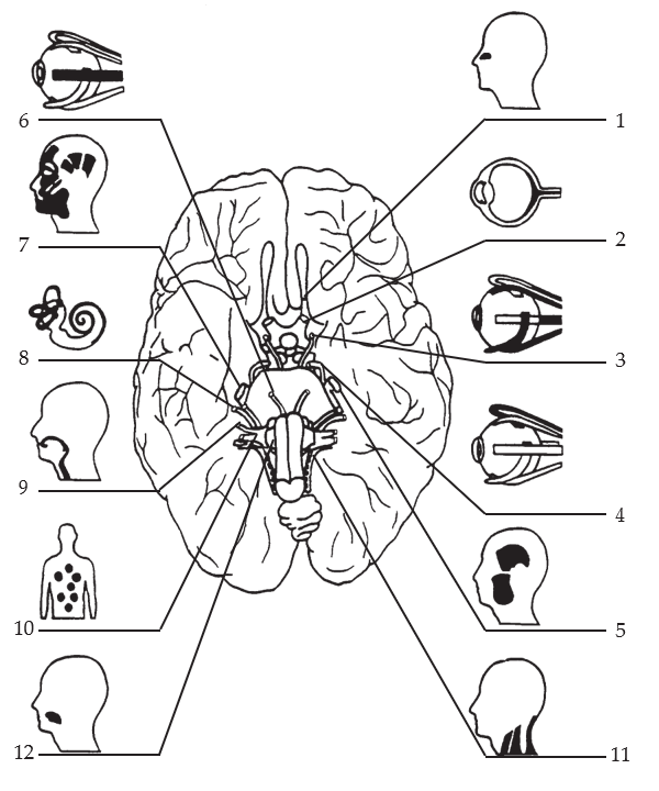
- State for each of the 12 pairs of cranial nerves, as given in Table 4.1 Functions of Parts of the Brain and Spinal Cord :
- sensory functions
- point of termination of sensory fibres in C.N.S.
- State for each of the 12 pairs of cranial nerves, as given in Figures 4.1, 4.2, and 4.3:
- point of origin of motor fibres in C.N.S.
- motor functions
Function Tables
Functions of Parts of the Brain and Spinal Cord
| Component of Brain | Cerebrum Integrative Functions | |
|---|---|---|
|
||
| Sensory Functions | Motor Functions | |
| Frontal lobe |
|
|
| Parietal lobe |
|
|
| Temporal lobe |
|
|
| Occipital lobe |
|
|
| Basal Nuclei | Assist with coordination of voluntary skeletal muscle movement by controlling associated subconscious movements which accompany voluntary activity (i.e. swinging arms when walking, facial expressions) | |
|
Brainstem (functions common to all parts of the brain stem) |
|
Reflex centre for cranial nerves to other parts of brain nerves (i.e. autonomic reflexes of salivary gland secretion, pupil dilation) |
|
Brainstem – Midbrain, Pons, and Medulla Oblongata |
Control Centre
|
|
|
Thalamus |
|
|
| Hypothalamus |
|
|
| Cerebellum
|
|
|
| Spinal Cord | Sensory projection tracts | Motor projection tracts
|
Sensory Functions of Cranial Nerves
| Cranial Nerves | Sensory Functions | In C.N.S. Termination Point | |
|---|---|---|---|
| I | OLFACTORY (Sensory) | Transmits impulses from olfactory receptors | Olfactory bulbs |
| II | OPTIC (Sensory) | Transmits impulses from retina. | Thalamus |
| V | TRIGEMINAL (Mixed) | 3 sensory branches: transmits impulses re: touch, pain, temperature from receptors in skin and mucous membranes of head, face, mouth, and tongue.
Proprioceptive sensory axons from the extrinsic eye muscles begin their course toward the brain in cranial nerves III, IV, and VI but enter the brainstem via the Trigeminal nerve. |
Pons |
| VII | FACIAL (Mixed) | Transmits impulses from taste receptors of anterior two-thirds of tongue. | Pons |
| VIII | VESTIBULOCOCHLEAR (Sensory) | 2 branches:
|
Pons and Medulla (Vestibule Nucleus) |
| IX | GLOSSO-PHARNGEAL (Mixed) | Transmits impulses from taste receptors of posterior one-third of tongue. Transmits impulse from baroreceptors and chemoreceptors in carotid sinus. | Medulla |
| X | VAGUS (Mixed) | Transmits impulses re: pressure, pain, proprioception from receptors in
RESPIRATORY ORGANS:
DIGESTIVE ORGANS:
HEART and AORTA |
Medulla Pons |
Somatic and Visceral Motor Functions of Cranial Nerves
(S) = Somatic Effector Response
(A) = Autonomic Effector Response
| Cranial Nerves | Motor Function | Point of Origin in C.N.S. | |
|---|---|---|---|
| III | OCULOMOTOR | Stimulates contraction of:
|
Midbrain |
| IV | TROCHLEAR | Stimulates contraction of extrinsic eyeball muscles – movement of eyeballs. (S) | Midbrain |
| V | TRIGEMINAL | Stimulates muscles of mastication for control of chewing movements. (S) | Pons |
| VI | ABDUCENS | Stimulates contraction of extrinsic eyeball muscles – movement of eyeball. (S) | Pons |
| VII | FACIAL | Stimulates:
|
Pons |
| IX | GLOSSO-PHARYNGEAL | Stimulates:
|
|
| X | VAGUS Somatic Fibres
|
Stimulates:
|
Medulla |
| VAGUS Autonomic Fibres | Stimulates:
Inhibits cardiac muscle contraction decreased rate and force of contraction. (A) |
||
| XI | ACCESSORY | Stimulates contraction of sternocleidomastoid and trapezius muscles → head movements. (S) | Spinal cord C1-5 (the fibres enter the cranium through the foramen magnum, then exit through the jugular foramen) |
| XII | HYPOGLOSSAL | Stimulates contraction of muscles of tongue → speech and swallowing. (S) | Medulla |
Sensory And Motor Functions Of The Cranial Nerves Summarized
(S) = Somatic Effector Response
(A) = Autonomic Effector Response
| Cranial Nerves | Sensory Functions | Motor Functions | |
|---|---|---|---|
| I | OLFACTORY | Transmits impulses from olfactory receptors | |
| II | OPTIC | Transmits impulses from retina. | |
| III | OCULOMOTOR | Stimulates contraction of:
|
|
| IV | TROCHLEAR | Stimulates contraction of extrinsic eyeball muscles – movement of eyeball. (S) | |
| V | TRIGEMINAL | 3 sensory branches: transmits impulses re: touch, pain, temperature from receptors in skin and mucous membranes of head, face, mouth, and tongue.
Proprioceptive sensory axons from the extrinsic eye muscles. |
Stimulates muscles of mastication for control of chewing movements. (S) |
| VI | ABDUCENS | Stimulates contraction of extrinsic eyeball muscles – movement of eyeball. (S) | |
| VII | FACIAL | Transmits impulses from taste receptors of anterior two-thirds of tongue. | Stimulates:
|
| VIII | VESTIBULO-COCHLEAR | Two branches:
|
|
| IX | GLOSSO-PHARYNGEAL | Transmits impulses from taste receptors of posterior one- third of tongue.
Transmits impulses from baroreceptors and chemoreceptors in carotid sinus. |
Stimulates:
|
| X | VAGUS | Transmits impulses re: pressure, pain, proprioception from receptors in:
RESPIRATORY ORGANS: DIGESTIVE ORGANS: HEART and AORTA |
Stimulates:
Inhibits cardiac muscle contraction → decreased rate and force of contraction (A) |
| XI | ACCESSORY | Stimulates contraction of sternocleidomastoid and trapezius muscles → head movements. (S) | |
| XII | HYPOGLOSSAL | Stimulates contraction of muscles of tongue → speech and swallowing. (S) | |
Lab Part 1: Parts of the Brain
In this lab activity, you will locate the parts of the brain using a model of the human brain and using a dissected sheep brain. This will also include stating the functions of the parts of the brain, and describing the production and flow of cerebrospinal fluid.
Materials Required:
- preserved sheep brains
- model of human brain
- model of fifth cervical vertebra
Part 1A: Brain Structure
- In the lab obtain a brain model and refer to the brain charts. Using the information from the learning objectives, locate and state the functions of the parts of the brain as indicated.
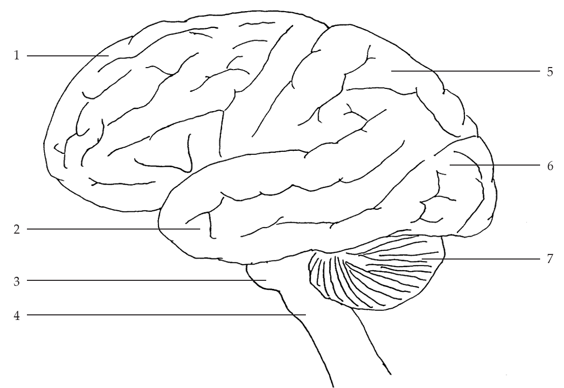
- Label Figure 4.1 Brain – Lateral View
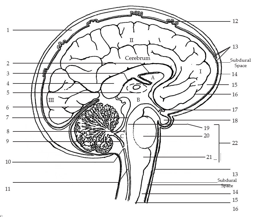
- Label Figure 4.2 Longitudinal Section Through Brain
Download and label the following figures by right-clicking on the image and choosing “Save Image As”.
Part 1B: Dissection of Sheep Brain
Note: there will be multiple sheep brains available in the lab; a whole brain, a brain cut in half longitudinally, and a brain cut in half transversly.
- Using the whole brain, position the brain with its ventral surface down in the dissecting tray, and locate the following features on the specimen.
- cerebral hemispheres
- longitudinal fissure
- frontal lobe }
- parietal lobe } of cerebrum
- occipital lobe }
- temporal lobe }
- cerebellum
- medulla oblongata
- convolutions/ridges (called gyri)
- shallow furrows (called sulci)
- deep furrows (called fissures)
- Bend the cerebellum and medulla oblongata slightly downward and away from the cerebrum. This will expose the pineal gland in the upper midline.
Note: The pineal gland secretes the hormone melatonin. In humans this contributes to the control of daily cycles of sleep and wakefulness. In sheep it is much larger relative to the size of the brain because it also controls the annual breeding cycle.
- With the brain in the same position, separate the cerebral hemispheres with your fingers until you can see the white piece of tissue connecting the two hemispheres.
Question: What is this white piece of nerve tissue called?
- Turn the specimen over and locate the parts of the brain listed below on its undersurface:
- cerebellum
- spinal cord
- occipital lobe
- medulla oblongata
- temporal lobes
- pons
- frontal lobe
- longitudinal fissure
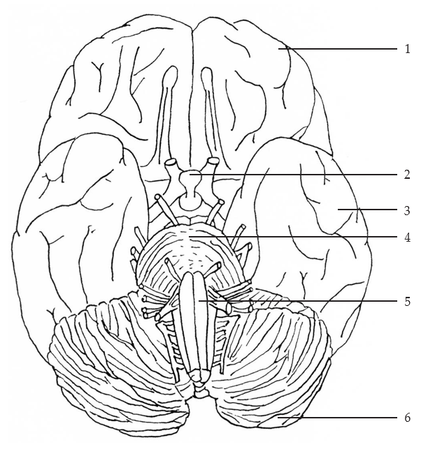
- Download and label Figure 4.3 Brain – Inferior Surface by right-clicking on the image and choosing “Save Image As”.
-
Using the transversely and longitudinally cut brains locate the structures listed below. On each of these dissected brains find as many of the structures as possible.
- cerebrum
- frontal lobe of cerebrum
- parietal lobe of cerebrum
- occipital lobe of cerebrum
- temporal lobe of cerebrum
- cerebellum
- corpus callosum
- medulla oblongata
- midbrain
- pons
- thalamus
- hypothalamus
- On the transversely cut brain locate the white and gray matter of the cerebrum.
Question: What forms the white and grey matter?
White –
Gray –
Question: Why is the white and grey matter arranged in this manner? (Grey on cortex, white matter central with fingers extending into grey).
Lab Part 2: Ventricles, Meninges and Cerebrospinal Fluid
In this lab activity you will locate the membranes and spaces surrounding the brain and spinal cord trace the production and movement of cerebrospinal fluid around the brain and spinal cord,
Materials Required
- dissected brain
- brain model
- ventricle model
- brain chart
- model of the 5th cervical vertebra
Part 2A – Ventricles
- Using the following materials available in the lab locate the lateral ventricles, and the 3rd and 4th ventricles:
- dissected brain
- brain model
- ventricle model
- brain chart
Part 2B – Meninges
Meninges and the Brain
- Examine then whole preserved brain and see if you can locate the 3 meninges listed below:
- Dura Mater – the thick, opaque outer layer.
- Arachnoid Mater – the delicate, transparent middle layer that is attached to the undersurface of the Dura Mater.
- Pia Mater – the thin, vascular layer that adheres to the brain.
Question: Where is the subarachnoid space? What does it contain?
Question: Where is the subdural space? What does it contain?
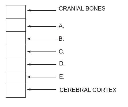
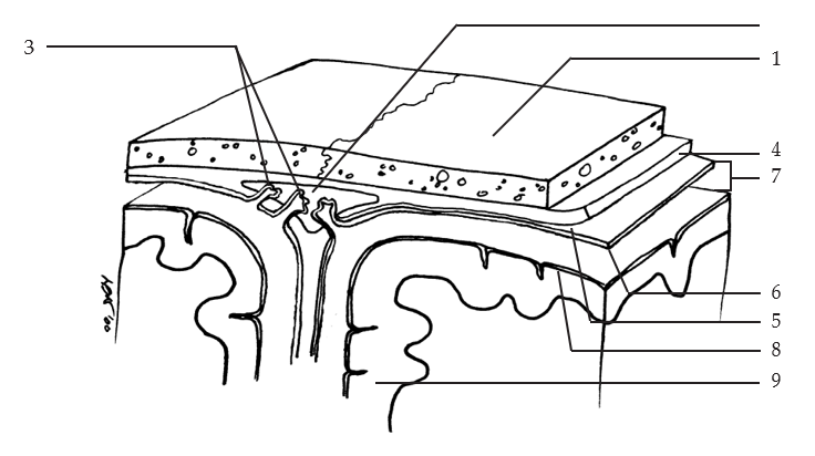
- Summarize your knowledge of the locations of the layers and spaces which surround the brain and spinal cord by completing Figure 4.4 Layers and Spaces Surrounding the Brain and Spinal Cord. Download and label the following figure by right-clicking on the image and choosing “Save Image As”.
- Download and label figure Figure 4.5 The Meninges by right-clicking on the image and choosing “Save Image As”.
Meninges and the Spinal Cord
- Using the model of the 5th cervical vertebra, locate the membranes and spaces surrounding the spinal cord.
- The inner layer adhering to the spinal cord is the [blank].
- The middle layer loosely covering the spinal cord is the [blank].
- The outer fibrous membrane is the [blank].
Question: Where is the epidural space? What does it contain?
Part 2C – Cerebrospinal Fluid
In an adult 80-150 ml is present in the ventricles and subarachnoid spaces. In a newborn 10 – 30 ml is present.
It is under pressure equal to a column of water 110 mm high. It is derived from the blood plasma and includes:
- water
- protein (15-40 mg/ml)
- white blood cells (less than 10 lymphocytes and monocytes/ml)
- electrolytes
- glucose (60-70% of blood plasma glucose)
- Trace the circulation of the C.S.F. through the brain and around the brain and spinal cord on the diagram provided in the lab. Include in your description the origin/production, pathway, and eventual removal of C.S.F.
Lab Part 3: Cranial Nerves
Materials Required:
- preserved sheep brain
Part 3A
- On the preserved sheep brain available in the lab, locate as many of the cranial nerves listed below in figure 4.6 Cranial Nerves as possible.
- olfactory bulb
- optic nerves
- optic chiasma
- optic tract
- oculomotor nerve
- trochlear nerve
- trigeminal nerve
- facial nerve
- vestibulocochlear nerve
- glossopharyngeal nerve
- vagus nerve
- (spinal) accessory nerve
- hypoglossal
- abducens
- Label the diagram (Fig.E) Brain with Cerebellum and Part of Left Cerebral Hemisphere removed. Download and label the following figure by right-clicking on the image and choosing “Save Image As”.
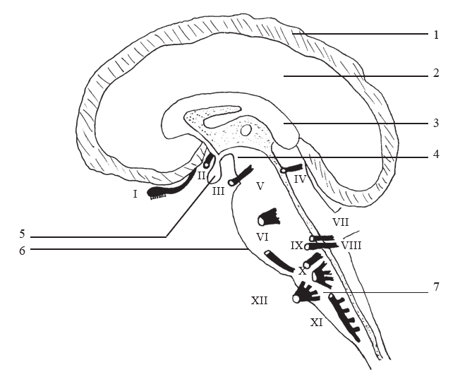
4.7 Brain with Cerebellum and Part of Left Hemisphere Removed
Practice: Names and Functions of the Cranial Nerves
Complete the following table, identifying the movements that result from stimulation of visceral and somatic effects by each cranial nerve.
Download Names and Functions of the Cranial Nerves Practice [DOCX] to print and fill out.
| Cranial Motor Nerves | Specific Names of Nerves | Movement Resulting From Control by Cranial Nerves |
|---|---|---|
| 1. Cranial Nerve III | [Blank] Nerve | Regulates [blank] size, [blank] movement and [blank] of the lens. |
| 2. Cranial Nerve IV | [Blank] Nerve | Regulates [blank] movements. |
| 3. Cranial Nerve V | [Blank] Nerve | Regulates [blank] mvoements. |
| 4. Cranial Nerve VI | [Blank] Nerve | Produces [blank] movements of the eye. |
| 5. Cranial Nerve VII | [Blank] Nerve | Produces expression of the [blank] and secretion of [blank] and tears. |
| 6. Cranial Nerve XI | [Blank] Nerve |
Controls [blank] movement, [blank] of head, and voluntary swallowing movement. |
| 7. Cranial Nerve XII | [Blank] Nerve | Produces [blank] movements involved in speech and swallowing activities. |
| 8. Cranial Nerve IX | [Blank] Nerve | Causes [blank] movements and secretion of [blank]. |
| 9. Cranial Nerve X | [Blank] Nerve | Causes movement of muscles in the [blank], [blank], and thoracic and [blank] viscera – i.e. [blank]of heart, [blank] Peristalsis in [blank] and [blank] digestive gland secretion. |
