
Labs and Activities
Week 1 Package 1 – Organs Of The Urinary System: Acroscopic Structure And Function
In this package, the student will study the macroscopic structure and function of the kidney, urinary bladder, ureters and urethra. This knowledge will serve as a basis for understanding the maladaptations that occur when the urinary system fails to perform its normal functions.
Learning Objectives
Upon completion of this learning package, the student will be expected to recall the following organs of the urinary system and their general function(s) in maintaining physiological integrity.
Click the triangle drop-down to see specific objectives:
Kidneys
- State the size and shape of the kidneys.
- Describe the location of the kidneys considering:
- their relationship to the parietal peritoneum and vertebral column (retroperitoneal position)
- the position of the right kidney in relation to the left kidney
- State, for each of the following features of the external structure of the kidney:
- renal capsule
- adipose capsule
- renal fascia
- renal hilum
- a definition of each component
- tissue makeup (where appropriate)
- its specific function(s)
- Identify, the location of each of the following macroscopic structures of the kidney:
- renal cortex
- renal columns
- renal medulla
- renal pyramids (renal papilla also)
- calyx (minor and major)
- renal pelvis
- renal sinus
- Recognize that the kidneys assist in the maintenance of homeostasis by:
- elimination of wastes
- maintenance of fluid and electrolyte balance
- maintenance of acid-base balance
- regulation of arterial blood pressure
- secretion of the hormone erythropoietin
Ureters
- Describe the ureters considering:
- their location in relation to the parietal peritoneum, kidney and bladder
- the three coats composing the walls and their respective functions.
- the significance of their oblique attachment to the bladder
- State one function of the ureters.
Urinary Bladder
- Describe the urinary bladder considering:
- its location in relation to the symphysis pubis
- the type, location and function of the three coats that make up the wall of the urinary bladder
- the three openings of the bladder
- a definition of “trigone”
- State two functions of the urinary bladder.
Urethra
- Describe the urethra considering:
- the type of membrane lining the urethra
- the location relative to the pelvic floor
- muscle tissue and type of control of the internal and external sphincters
- the location and function of the urinary meatus (external urethral orifice)
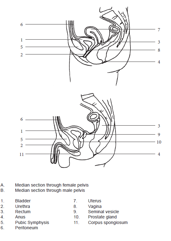
- Differentiate between the male and female urethra considering:
- the anatomical location and length
- the specific function(s) of each
Lab Activity: Components Of The Urinary System And Dissection Of The Pig Kidney
This lab activity is designed to provide experience locating and describing the structures of the urinary system through the use of a kidney model and laboratory dissection of a pig kidney.
Materials Required:
- Human Torso model
- Urinary system model
- Dissecting tray and kit
- Fresh pig kidney
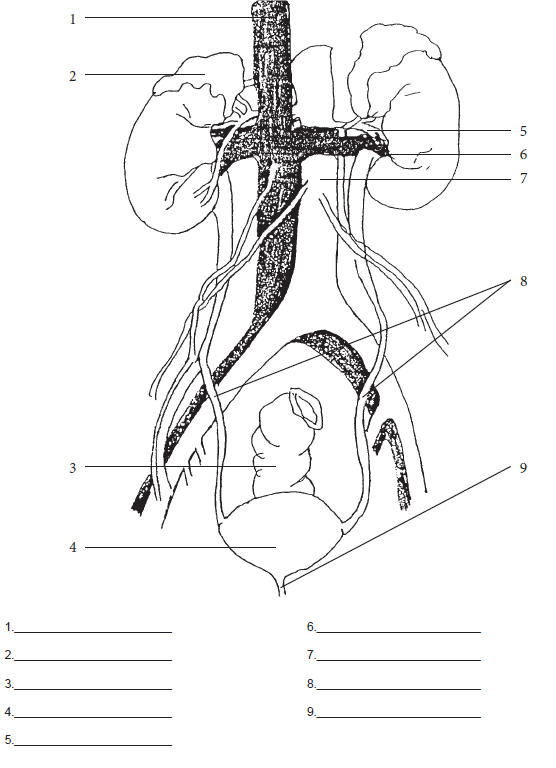
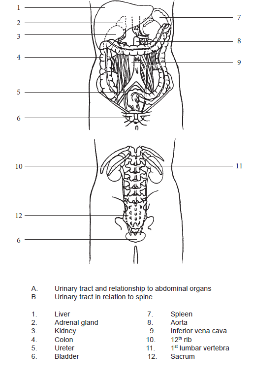
Part A – Organs of the Urinary System
- Use the model of the human torso and the model of the urinary system. Locate and describe the following structures:
- Kidneys
- Ureters
- Urinary Bladder
- Urethra
- Download and label the following figures by right-clicking on the image and choosing “Save Image As”.
Part B – Dissection of the Pig Kidney
External Surface
- Before cutting into the kidney, observe the amount of fat encasing it (if present).
- Look for a small gland (adrenal gland) embedded in the fat around the superior end of the kidney.
- Starting at the convex border of the kidney, carefully peel the fat from around the kidney, using care not to tear away the vessels that enter the kidney.
- About 1” from the hilum (this is the area/depression where blood vessels, lymphatics and nerves enter/leave an organ), cut the fat off cleanly, using a scalpel blade. Be careful not to damage any vessels.
Question: What is the function of the fat surrounding the kidney?
- Locate the following components on your specimen:
- renal capsule
- hilum
- renal artery
- renal vein
- ureter
- Examine the shiny, transparent covering membrane (if present), adhering to the kidney. Remove a piece of it with your forceps. State the name of the components of the external structure of the kidney and state their functions.
- Carefully insert a probe into the renal artery and vein.
Question: How far can your probe enter the kidney?
The Ureter
- Carefully insert your probe into the ureter and leave it in place.
Question: What is the function of the ureter?
Question: What are the names of the three layers that make up the wall of the ureter?
- Describe the tissues that make up the wall of the ureter, along with their functions.
Internal Structure
- Using a sharp scalpel, cut the kidney in half longitudinally along the frontal plane. (Ask for instructions if you are not sure how to do this).
- Separate the two halves completely. Lay the open organ on the dissecting tray. Locate the tip of the probe that was placed in the ureter.
Question: What part of the kidney does the ureter open into?
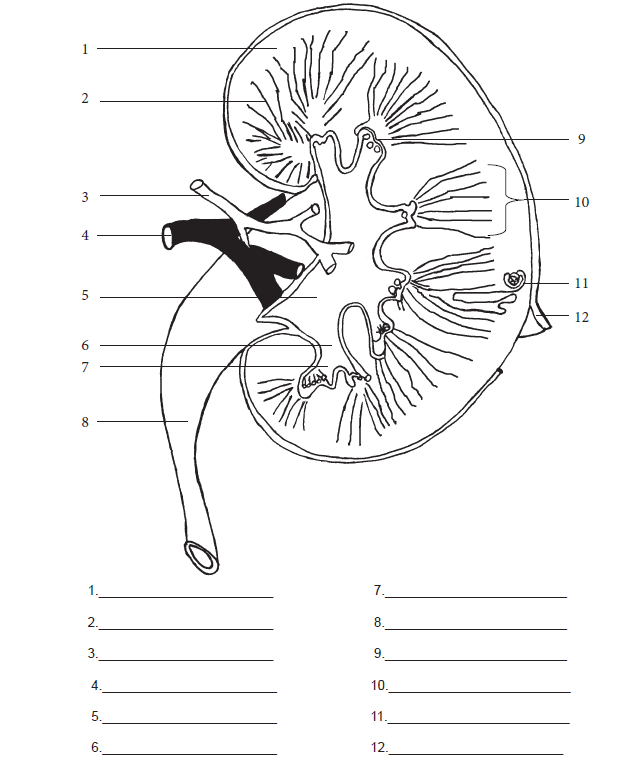
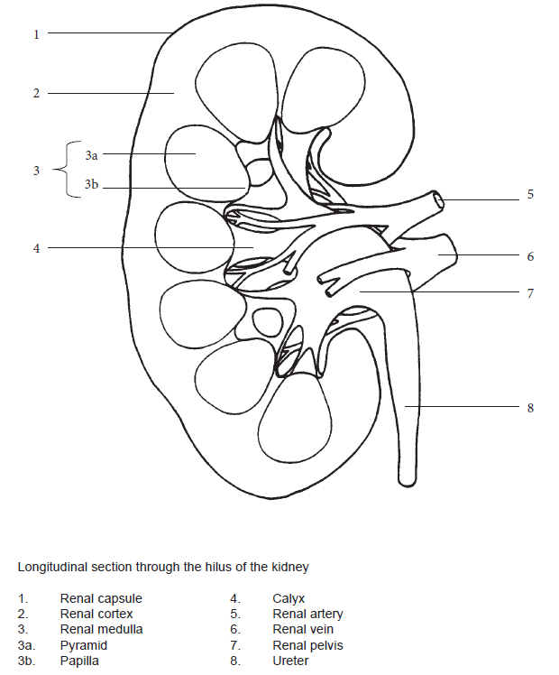
- On your dissected kidney locate the following structures:
- renal capsule
- renal papilla
- renal cortex
- renal pelvis
- renal medulla
- renal calyces
- renal pyramid
- renal columns
- Label the structures on the diagrams below.
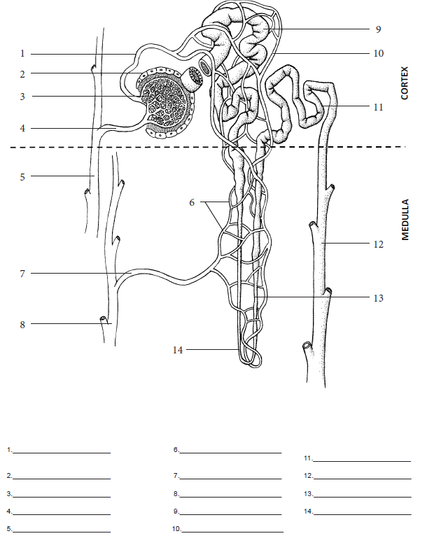
- Trace an imaginary nephron on the dissected kidney.
- Describe the appearance of the cortex. List the parts of the nephron and blood vessels that make up the cortex.
- Describe the appearance of the medulla.
- List the parts of the nephron and blood vessels that make up:
- the renal pyramids.
- the renal columns.
- Trace the path of the filtrate from the Bowman’s capsule to the urethra using diagrams and the kidney specimen.
- Trace the path of blood flow from the renal artery to renal vein on your dissected kidney specimen.
Once you have completed your objectives, questions, and diagrams, do your evaluation. When you have completed your evaluation, throw out the kidney in the Biohazard container and wash the equipment.
