
Labs and Activities
Week 7 – White Blood Cells, Platelets, and Lymphatic System
Learning Objectives
Upon completion of this learning package, the student will be expected to recall the following.
Click the triangle drop-down to see specific objectives:
White Blood Cells
- Describe white blood cells (leukocytes) as follows:
- site of formation
- general function
- classification according o two main categories
- name and function of each specific style of white blood cell
Platelets
- Describe platelets (thrombocytes) as follows:
- site of formation
- general function
Lymphatic System
- State the three main functions of the lymphatic system as given below:
- return of excess tissue fluid, protein and fat to the blood via lymphatic
- vessels
- production of lymphocytes, monocytes and plasma cells for body defense by lymph nodes
- transport of lipids following absorption from small intestine
- Describe the path followed by the fluid that enters the lymphatic vessels until it exits into the blood using the following guidelines:
- source of the excess ISF in all tissues
- role of lymphatic capillaries
- role of smaller lymph vessels
- role of five principal lymphatic trunks
- names of two main lymphatic vessels into which the lymph trunks empty
- parts of the body drained by each main lymphatic vessel identified in e)
- site where each main lymphatic vessel empties into the blood
- Describe how lymph vessels differ from blood vessels in terms of the following:
- description of capillary walls
- three ways lymph vessels differ from veins
- source of the pressure which moves the lymph
- whether the system is open or closed
- Lymph passes through lymph nodes as it moves along lymph vessels to be returned to the circulatory system. State the function(s) of lymph nodes. Locate on a diagram – five main groups of lymph nodes listed below:
- submental and submandibular groups in the floor of the mouth
- cervical group along the sternocleidomastoid muscle in the neck
- supratrochlear group above the bend in the elbow
- axillary group within the underarm and upper chest
- inguinal group in the groin
- Outline briefly and show, using a diagram of the lymph node, each of the following:
- the structure, including the capsule, trabeculae, cortex, medulla, medullary cords, afferent lymphatic vessels, sinuses, efferent lymphatic vessels.
- the mechanics of lymph flow through the node
- the types of white blood cells found within the node
- the type of phagocytic cells found within the node
- its hemopoietic functions
- its role in specific immune response
- Distinguish between a lymph node and a lymph nodule in terms of structure, components and fluid screened by the structure(s).
- Describe a lymph nodule and locate on a diagram, the following specific examples of lymph nodules or mucosa-associated lymphatic tissue (MALT):
- palatine tonsils
- lingual tonsils
- pharyngeal tonsil (adenoid)
- Peyer’s patches
- Describe the structure and function of the lymphatic organ, the thymus gland, according to the following criteria:
- location, in relation to the trachea and sternum
- period in life cycle when it is most functional
- role in specific immunity
- Describe the structure and function of the lymphatic organ, the spleen, according to the following criteria:
- location, in relation to diaphragm and stomach
- constituents of the stroma, white pulp and red pulp
- two protective functions with regard to:
- hemopoiesis
- phagocytosis
Lab Activities
It is usually necessary to know not only the total number of white blood cells but also the relative abundance of each leukocyte type. This knowledge is obtained by determining the number of each leukocyte type out of a total count of 100 white blood cells.
Materials Required:
- compound microscope
- prepared blood slide
Part A – Differential White Blood Cell Count
- Review the appearance of red blood cells, platelets and the white blood cells listed in the table below by referring to diagrams in your text book and posters available in the lab.
- Place the blood slide on the stage of the microscope and focus at 400x.
Question: How is the appearance of the white blood cells different from the appearance of the red blood cells?
- In the first field of view how many white blood cells do you see? Oh these, how many are neutrophil? Eosinophils? Basophils? Monocytes? Lymphocytes?
- Use the counter to record the total number of white blood cells that you see. Record the number of each type in the table.
- Now move the slide to the right or left until a new field of view is in place. Again, count the total number of white blood cells and record the number of each type of cell in the table as you did in step 4.
- Again, move the slide to a new field of view and record the number of white blood cells and the number of each type. Repeat this process until you come to the edge of the coverslip.
- When you reach the edge, move the slide up or down until a new field of view is in place. Again, do your counts and then move across the slide as you did before. Refer to the diagram available in the lab that will show the pattern of movement back and forth across the slide.
- When the counter reaches 100, stop counting.
- For each type of cell calculate the percentage of the total number of cells that you have counted and record this value in the table.
- Now obtain a slide of monocytic leukemia and TRY to do the same count as you did for the normal slide.
| Cell Type | Cell Count – Normal Slide | Percentage % | Cell Count – Leukemia Slide | Percentage % |
|---|---|---|---|---|
| Neutrophils | ||||
| Eosinophils | ||||
| Basophils | ||||
| Lymphocytes | ||||
| Monocytes |
Question: How do the percentages of each cell type that you have counted compare with the normal values?
Question: Describe how the leukemia slide is different from the normal slide. How might these differences make it hard to do a differential white blood cell count?
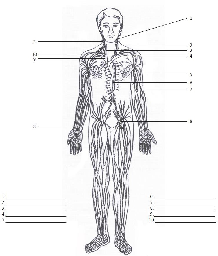
Part B – Lymphatic System: Location of Vessels and Nodes
- Locate the following structures on figure 4.2 The Lymphatic System
- Blood vessels
- Internal jugular veins (left and right)
- Right subclavian vein
- Left subclavian vein
- Lymph vessels
- Thoracic duct
- Right lymphatic duct
- Lymph nodes
- Submaxillary group
- Cervical group
- Axillary group
- Supratrochlear group
- Inguinal group
- Blood vessels
- For each component listed below in Column 1 (1-11), select its location from the alternatives in Column 2 (A-K)
| Column 1 – Components | Column 2 – Locations |
|---|---|
| 1. Peyer’s patches | A. In groins, draining external genitalia, and legs. |
| 2. Spleen | B. Below and behind pillars of fauces. |
| 3. Adenoids | C. In neck, along sternocleidomastoid muscle. |
| 4. Lingual tonsils | D. at the base of the tongue. |
| 5. Palatine tonsils | E. In the (L) hypochondrium, above (L) kidney, below the diaphragm. |
| 6. Axillary lymph nodes | F. Mucous membrane layer of small intestine. |
| 7. Cervical lymph nodes | G. Within underarm and upper chest regions. |
| 8. Supratrochlear (cubital) lymph nodes | H. In the mediastinum, extending between the 4th costal cartilage and lower edge of thyroid. |
| 9. Submental, and submaxillary nodes | I. In nasopharynx, on posterial wall. |
| 10. Inguinal nodes | J. Just above bend in elbow. |
| 11. Thymus gland | K. In the floor of the mouth, draining the nose, lips and teeth. |
